H-3000 Hatvan, 17 Kisfaludy Street
  +36 37 999 650
  informacio@mc60.hu
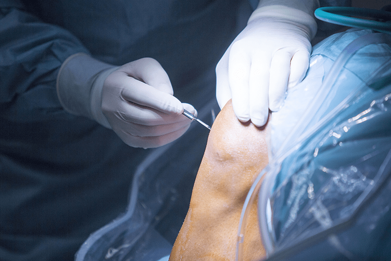
Arthroscopy, also known as joint arthroscopy, is a minimally invasive surgical procedure most used to diagnose and treat joint problems, such as those affecting the knee, shoulder, hip or ankle.
During the procedure, the surgeon makes a small incision in the skin through which an arthroscope is inserted into the joint. Using a camera, the surgeon obtains a detailed view of the inside of the joint and its condition. This makes it possible to identify and diagnose a wide range of abnormalities and, where necessary, to treat them during the same procedure.
Although arthroscopy is less invasive than traditional open surgery, it is still a significant surgical procedure. However, it is generally associated with a faster recovery and less postoperative pain for the patient.
Arthroscopy and rotator cuff repair is a minimally invasive shoulder procedure used to treat injuries of the shoulder joint, particularly rotator cuff tears. During the operation, the surgeon inserts a camera and specialised instruments into the joint through small incisions, allowing the damaged tendons to be accurately assessed and treated. The aim of rotator cuff repair is to reattach the torn or damaged tendon to the bone, restoring shoulder stability, range of motion and pain-free function. Before the procedure, a thorough orthopaedic assessment and adherence to your surgeon's instructions are essential. Following surgery, protecting the shoulder, attending physiotherapy and following the prescribed rehabilitation programme are key to a successful recovery.
Arthroscopic debridement is a minimally invasive joint procedure in which the surgeon uses a specialised camera and surgical instruments to remove damaged, inflamed or dead tissue from within the joint. It is most performed to treat cartilage damage, degenerative joint disease, inflammation or sports injuries to relieve pain and improve joint mobility. The procedure is carried out through small incisions and may therefore result in less tissue trauma and a faster recovery than traditional open surgery. Before the operation, a comprehensive orthopaedic assessment and compliance with medical advice are important, while after surgery, rest, physiotherapy and adherence to the prescribed rehabilitation programme play a vital role in achieving optimal healing.
Arthroscopic anterior cruciate ligament (ACL) reconstruction is a minimally invasive orthopaedic procedure most performed to treat injury or rupture of the anterior cruciate ligament of the knee. During the operation, the surgeon inserts a camera and specialised instruments through small incisions into the joint and reconstructs the damaged ligament using a graft taken either from the patient's own tendon or from donor tissue. The aim of the procedure is to restore knee stability, reduce pain and improve joint function, particularly in athletes and physically active individuals. A thorough orthopaedic assessment and careful adherence to medical advice are important before surgery. Following the procedure, rehabilitation, physiotherapy and the gradual return to weight-bearing activities are essential for a full recovery.
Arthroscopic meniscal surgery is a minimally invasive knee procedure performed to treat injuries to the meniscus within the knee joint. The meniscus is a resilient cartilage structure that plays an important role in knee stability and the distribution of load across the joint. During the operation, the surgeon inserts a camera and specialised instruments through small incisions, then removes the damaged portion of the meniscus or repairs it using sutures where appropriate. The aim of the procedure is to relieve pain, restore normal knee function and help prevent further damage to the joint. Before surgery, a thorough orthopaedic assessment and compliance with medical advice are essential. After the procedure, rest, physiotherapy and following the prescribed rehabilitation programme play an important role in ensuring a successful recovery.
Arthroscopic mosaicplasty is a minimally invasive orthopaedic procedure used to treat damage to the articular cartilage, most commonly in the knee joint. During the operation, the surgeon transplants small cylindrical plugs of healthy cartilage and bone from less weight-bearing areas to the damaged cartilage surface, helping to restore the joint's natural surface. The aim of the procedure is to reduce pain, improve joint function and slow the progression of cartilage wear. Thanks to the arthroscopic technique, the procedure may result in less tissue trauma and a faster recovery than conventional surgery. Before the operation, a thorough orthopaedic assessment and adherence to your surgeon's instructions are essential. Following the procedure, avoiding weight-bearing, attending physiotherapy and following the prescribed rehabilitation programme are key to achieving a successful recovery.
Arthroscopic ankle surgery is a minimally invasive orthopaedic procedure used to treat a variety of ankle joint injuries and conditions. During the operation, the surgeon inserts a camera and specialised instruments through small incisions, allowing cartilage injuries, inflammation, loose bodies and ligament injuries to be accurately identified and treated. The aim of the procedure is to relieve pain, improve joint mobility and restore ankle stability. Compared with traditional open surgery, the arthroscopic technique may result in less tissue trauma, smaller scars and a faster recovery. Before the operation, a thorough orthopaedic assessment and adherence to your surgeon's instructions are essential. After surgery, rest, gradual return to weight-bearing activities, physiotherapy and following the prescribed rehabilitation programme all play an important role in recovery.
Arthroscopic elbow surgery is a minimally invasive orthopaedic procedure used to treat a range of elbow joint injuries, inflammatory conditions and degenerative changes. During the operation, the surgeon inserts a camera and specialised instruments through small incisions, allowing cartilage injuries, adhesions, loose bodies and other joint problems to be accurately identified and treated. The aim of the procedure is to relieve pain, improve range of motion and restore normal joint function. Compared with traditional open surgery, the arthroscopic technique may result in less tissue trauma, a shorter recovery period and faster rehabilitation. Before the operation, a thorough orthopaedic assessment and adherence to your surgeon's instructions are essential. Following surgery, rest, physiotherapy and adherence to the prescribed rehabilitation programme are key to achieving a successful recovery.
Arthroscopic shoulder surgery with acromioplasty is a minimally invasive orthopaedic procedure most performed to treat shoulder impingement syndrome. In this condition, the tendons and soft tissues of the shoulder become compressed beneath the acromion, causing pain, restricted movement and inflammation. During the operation, the surgeon inserts a camera and specialised instruments through small incisions, removes inflamed tissue and, where necessary, reshapes the undersurface of the acromion to create more space for the tendons. The aim of the procedure is to relieve pain, improve shoulder movement and prevent further damage. Before the operation, a thorough orthopaedic assessment and adherence to your surgeon's instructions are essential. Following surgery, physiotherapy, gradual return to activity and adherence to the prescribed rehabilitation programme are vital for a successful recovery.
The Denks procedure is an orthopaedic knee operation performed primarily to treat abnormal positioning of the kneecap (patella), overuse, degenerative changes or recurrent patellar instability and dislocation. During the procedure, the bony attachment of the patellar tendon is repositioned together with a bone block and secured with a screw, improving patellar tracking and restoring normal knee function. The aim of the operation is to relieve pain, improve patellar stability and prevent further dislocations. Recovery typically involves the use of a knee brace, crutches, gradual weight-bearing and physiotherapy. Bone healing usually takes around six weeks, while rehabilitation often lasts approximately three months.
Bursitis surgery is performed when inflammation of the bursa is severe, chronic or has not responded to conservative treatment. During the procedure, the inflamed bursa is removed, although a new bursa may form naturally over time. The operation may be performed using either an open or a minimally invasive (arthroscopic) technique. Recovery usually takes several weeks, although returning to full weight-bearing activities may take longer.
Bunion surgery is a surgical procedure performed to correct a bunion, a deformity affecting the joint at the base of the big toe. A bunion is a bony prominence that develops at the big toe joint and can cause pain, inflammation and deviation of the toe. The aim of surgery is to relieve pain, realign the toe and restore normal foot function. Several surgical techniques are available depending on the severity of the deformity. Following surgery, patients usually wear a special rigid-soled post-operative shoe to protect the foot and support healing. Full recovery typically takes between 6 and 12 weeks, depending on the type of procedure performed and the individual's condition. Physiotherapy is often recommended to restore strength and mobility. Bunion surgery provides an effective solution for patients whose symptoms have not improved with conservative treatment.
Carpal tunnel release surgery is performed to relieve compression of the median nerve at the wrist. Pressure on the median nerve within the carpal tunnel can cause numbness, pain and weakness in the hand. During the procedure, the surgeon divides the transverse carpal ligament (transverse carpal ligament) to release pressure on the nerve. The operation may be performed using either an open or a minimally invasive endoscopic technique. It is usually carried out under local anaesthesia, although general anaesthesia may be appropriate in certain cases. Local anaesthesia is most used because it places less strain on the body and allows for a quicker recovery. Only the wrist and hand are numbed, allowing the patient to remain awake without feeling pain. General anaesthesia is rarely required but may be considered if the patient is particularly anxious or if the procedure is being performed alongside another operation. The decision is made jointly by the surgeon and the patient, considering the patient's overall health and individual circumstances. Recovery may take from several weeks to several months, with hand function improving gradually over time.
Cubital tunnel release surgery is a procedure performed to relieve pressure on the ulnar nerve at the elbow. The ulnar nerve passes through a narrow passage called the cubital tunnel, and when compressed it can cause numbness, pain and weakness in the forearm and hand, particularly affecting the ring and little fingers. During the operation, the surgeon relieves the pressure on the nerve by releasing it, repositioning it, or dividing the tissues that form the walls of the cubital tunnel. Recovery may take anywhere from several weeks to several months, and physiotherapy is often recommended to help restore full hand function.
Bone spur removal is an orthopaedic surgical procedure performed when a bony growth on the surface of a bone causes pain, restricted movement, difficulty wearing footwear, or irritation of nearby tendons, muscles or nerves, or when conservative treatment has not provided adequate relief. During the operation, the surgeon removes or reshapes the bone spur. Depending on its location, the procedure may be performed through an open approach or using minimally invasive arthroscopic techniques. If necessary, the removed tissue may be sent for histological examination. Recovery depends on the location of the bone spur and the extent of the procedure. Symptoms may improve within a few days or several weeks, while returning to full weight-bearing activities or work may take longer. In some cases, a brace, crutches and physiotherapy may also be required.
Wrist plate fixation is an orthopaedic trauma procedure most performed to treat fractures around the wrist, particularly fractures of the distal radius. During the operation, the fractured bone fragments are realigned into their anatomical position and stabilised using a specially designed metal plate and screws to ensure proper bone healing. The aim of the procedure is to restore wrist stability, prevent deformity and enable the hand to regain normal function as quickly as possible. Temporary immobilisation, gradual return to weight-bearing activities and physiotherapy may be required after surgery. Bone healing usually takes 6–8 weeks, while full recovery of movement and function may take several months.
Dupuytren's contracture is a chronic connective tissue disorder affecting the fibrous layer beneath the skin of the palm. As the condition progresses, scar-like cords develop in the palm and gradually shorten, pulling the fingers—most commonly the ring and little fingers—towards the palm. As a result, the fingers cannot be fully straightened, which can interfere with normal hand function. The exact cause is unknown, although genetic predisposition, diabetes, alcohol consumption and other factors may contribute to its development. Treatment is not always required in the early stages, but more advanced cases may require injection therapy, physiotherapy or surgery to restore finger movement.
A fémeltávolítás egy ortopédiai vagy traumatológiai műtét, amelynek során korábbi törésrögzítéshez vagy egyéb csontsebészeti beavatkozáshoz beültetett fémimplantátumokat – például lemezeket, csavarokat, szegeket vagy drótokat – távolítanak el. A műtétre általában akkor kerül sor, ha a csont teljesen meggyógyult, illetve, ha az implantátum fájdalmat, irritációt, mozgáskorlátozottságot vagy egyéb panaszt okoz. A beavatkozás során a korábbi műtéti hegen keresztül feltárják az érintett területet, majd eltávolítják a rögzítő eszközöket. A gyógyulás rendszerint néhány hétig tart, azonban a teljes terhelhetőség és a végleges funkcionális felépülés a műtét helyétől és kiterjedésétől függően hosszabb időt is igénybe vehet. Szükség esetén a rehabilitáció részeként gyógytorna javasolt.
Ganglion cyst removal is a surgical procedure performed to remove a benign, fluid-filled cyst that most commonly develops on the wrist, hand or foot. Although ganglion cysts are usually harmless, surgical removal may be recommended if they cause pain, restricted movement or cosmetic concerns. The procedure may be carried out using either an open or arthroscopic technique and aims to remove not only the cyst itself but also the associated joint capsule or tendon sheath from which it arises, reducing the risk of recurrence. Recovery usually takes several weeks, and gradual return to normal use of the wrist or affected area is recommended.
Reconstruction of the extensor pollicis longus (EPL) tendon is performed when the tendon has ruptured, been injured or has degenerated, resulting in significant limitation of thumb extension. During the procedure, the surgeon either repairs the tendon directly using sutures or reconstructs it by transferring another tendon, most commonly the extensor indicis proprius (EIP). Following the operation, the hand is immobilised, and physiotherapy is required to restore normal movement and function. Recovery usually takes between 6 and 12 weeks, although regaining full strength and range of motion may take longer.
Foreign body removal is a surgical procedure performed when a foreign object—such as glass, metal, wood or other fragments—has become embedded in the body and causes pain, inflammation, infection, restricted movement or other symptoms, or cannot be removed naturally. During the operation, the surgeon exposes the affected area, identifies and removes the foreign body, using imaging techniques such as X-ray or ultrasound if necessary. Depending on the size and location of the foreign body, the procedure may be performed under local, regional or general anaesthesia. Recovery usually takes from a few days to several weeks, with the overall recovery time depending on the condition of the surrounding tissues and the extent of the procedure. Where appropriate, wound care, antibiotic therapy or physiotherapy may also be required.
Biceps tendon repair is performed when the biceps tendon has partially or completely ruptured, causing pain, weakness and restricted movement of the arm. During the operation, the torn tendon is reattached to the bone—most commonly at the shoulder or elbow—using specialised sutures, anchors or other fixation devices. The procedure may be performed as an open operation or using minimally invasive arthroscopic techniques. Recovery typically takes 3–6 months, and physiotherapy is essential to restore full strength and range of motion.
Tendon repair surgery on the hand is indicated when a tendon in the hand has ruptured, for example because of an accident or injury. Tendons enable movement between the muscles and bones, so if a tendon is damaged, surgery is required to restore movement and function of the affected fingers. During the operation, the doctor repositions and sutures the ends of the torn tendon so that it can function fully again. The procedure may be performed as open surgery or arthroscopically, depending on the location of the tendon. Recovery time is usually 6–12 weeks. Rehabilitation, physiotherapy and gradual loading are required to regain hand function.
The Broström-Gould procedure is a specialised surgical procedure used to treat chronic instability of the lateral ankle ligaments. This operation is primarily recommended for patients who experience recurrent ankle sprains or ankle instability, and for whom conservative treatments, such as physiotherapy or the use of orthoses, have not produced adequate results. During the Broström technique, the surgeon tightens and secures the ligaments with sutures in their original position, thereby restoring stability to the ankle joint. The Gould technique is the second part of the procedure, during which the surgeon provides additional stability by using tendon and soft-tissue structures to further reinforce the ligaments. This provides a form of internal support for the ligaments. The Broström-Gould procedure is an effective solution for treating chronic ankle instability, particularly in patients for whom conservative therapies have not been successful.
Hammer toe deformity correction surgery is an orthopaedic procedure performed when hammer toe develops, where the middle joint of the toe becomes fixed in a bent position, causing pain, difficulty walking, calluses or problems wearing shoes. The aim of the operation is to correct the deformity, straighten the toe and reduce pain. During the procedure, the surgeon corrects the affected joint, tendons or bones, if necessary, by removing part of the bone or using temporary fixation. The operation may be performed using a traditional or minimally invasive technique. Recovery usually takes 4–8 weeks, during which wearing a special offloading shoe and gradual weight-bearing are recommended. Full functional recovery and resolution of swelling may take several months.
Trigger finger surgery is required when the symptoms of trigger finger cannot be relieved with conservative treatments such as rest, anti-inflammatory medication or injections. Trigger finger is a condition in which the tendons in the fingers catch within a narrow tendon sheath, resulting in painful clicking and restricted movement. During the operation, the surgeon makes a small incision in the finger and releases the tight band of connective tissue around the tendon that prevents it from moving freely. This helps resolve the trigger finger problem and allows the fingers to move freely. The operation is usually performed under local anaesthesia, and recovery time is 1–2 weeks, although full rehabilitation, including regaining movement and rebuilding strength, may take several months. The procedure may also be performed under general anaesthesia, but this is rarely necessary. It may be justified if the patient is very anxious or if it is combined with another operation. The decision regarding general anaesthesia is made jointly by the doctor and the patient.
Percutaneous pinning is a minimally invasive orthopaedic surgical procedure used primarily to stabilise certain bone fractures. During the procedure, the surgeon inserts special metal pins or wires into the bone through small skin incisions to hold the fractured bone fragments in the correct position and promote healing. The advantage of this method is that it involves less tissue trauma, may reduce the size of surgical scars and may allow faster rehabilitation. Before the operation, a detailed orthopaedic assessment and adherence to medical instructions are important, while after the procedure, follow-up appointments, appropriate immobilisation and compliance with instructions regarding gradual weight-bearing and physiotherapy play a key role.
Tarsal tunnel syndrome (TTS) involves compression of the tibial nerve and its branches, which may cause pain, numbness and weakness in the foot and ankle. The aim of tarsal tunnel release surgery is to reduce pressure on the nerve, thereby relieving the symptoms. During the procedure, the surgeon releases the nerve from compression so that blood circulation and nerve function can be restored. Tarsal tunnel release surgery can effectively reduce or eliminate the symptoms of tarsal tunnel syndrome, particularly when the diagnosis is made at an early stage.
Tennis elbow surgery is required when conservative treatments, such as rest, anti-inflammatory medication and physiotherapy, do not provide lasting improvement, and pain and restricted movement persist. Tennis elbow, or lateral epicondylitis, refers to inflammation of the tendons on the outer side of the elbow, most caused by repetitive strain. During the operation, the surgeon removes part of the damaged tendon or reconstructs it surgically to eliminate pain and restore elbow function. The procedure may be performed as open surgery or using an arthroscopic technique, where the operation is carried out through smaller incisions. Recovery time may be 6–12 weeks, depending on the type of surgery, and rehabilitation is required to regain full elbow function.
Removal of a pinning wire or screw is a surgical procedure performed when a device used to fix a bone fracture, such as a pinning wire or screw, is no longer needed, or if the fixation device causes pain or irritation. The procedure is usually performed under local anaesthesia so that the patient does not feel pain while remaining awake. The surgeon makes a small incision in the skin and then removes the pinning wire or screw. The operation is quick and can often be performed as an outpatient procedure. Recovery time is short, and the patient can usually return to everyday activities on the day of surgery. However, rest and follow-up appointments may be required to monitor post-operative scarring and complete bone healing.
Our institution’s musculoskeletal surgeon:
Contact us!