An ultrasound examination is a safe and painless imaging procedure that uses sound waves to examine the internal parts of the body. The sound waves are reflected from organs and tissues, and images are created from these returning signals, which the physician can evaluate. The examination does not use radiation, therefore ultrasound can be repeated and is risk-free at any age, whether for a child, an adult, or a pregnant woman. Ultrasound helps reveal the size, shape, and structure of organs, as well as any possible fluid accumulations, cysts, tumours, or inflammatory changes.
An ultrasound examination may be needed when your doctor wants to check the condition of internal organs or tissues. Common reasons include:
In certain situations, ultrasound may not provide reliable results, or the examination may need to be postponed. Such cases may include:
This examination assesses the condition of muscles, tendons, fatty tissue (e.g. lipoma), lymph nodes, and other soft tissues. It helps precisely identify injuries, inflammation, or benign lesions.
Preparation:
This examination evaluates the internal organs of infants and the development of the hip joints. It helps detect abnormalities early, such as hip dysplasia or abdominal organ disorders, allowing timely intervention.
When can the examination be performed?
Preparation:
This examination evaluates the condition of the testes, epididymis, and lymphatic regions. It helps detect inflammation, cysts, tumours, or other abnormalities at an early stage, enabling timely treatment.
Preparation:
This examination evaluates the abdominal organs (liver, gallbladder, kidneys, spleen, pancreas) and pelvic organs (urinary bladder, female or male reproductive organs). It helps detect inflammation, cysts, tumors, stones, or other abnormalities early, allowing prompt treatment.
Preparation:
This examination assesses multiple regions at the same time: axillary (armpit), abdominal, inguinal (groin), pelvic, and cervical (neck). It supports the early detection of tumors, enlarged lymph nodes, or other pathological processes, allowing timely initiation of appropriate treatment.
Preparation:
This examination evaluates the soft tissues of the neck and the main cervical arteries (carotid arteries). Using Doppler technology, not only the structure of the vessels but also the speed and direction of blood flow can be assessed. It helps detect narrowing, calcification, or circulatory disorders that may increase the risk of stroke.
Preparation:
This examination evaluates the thyroid gland, surrounding neck soft tissues, and lymph nodes. It helps detect nodules, inflammatory changes, cysts, or other abnormalities. It plays an important role in investigating thyroid dysfunction and monitoring known conditions.
Preparation:
Knee ultrasound evaluates the soft tissues around the joint – tendons, ligaments, muscles, joint capsule, and fluid collections. It helps detect injuries, inflammation, tears, or fluid accumulation. It is particularly useful in cases of sports injuries, pain, swelling, or limited mobility.
Preparation:
This procedure allows the doctor to precisely locate a thyroid nodule or other abnormality using ultrasound, and then collect a cell sample with a thin needle. The sample is examined in a laboratory, enabling a more accurate diagnosis of thyroid nodules, cystic lesions, or other soft tissue abnormalities. The procedure is quick, usually causes only mild discomfort, and is performed under local anesthesia.
Preparation:
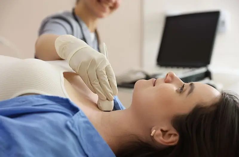
Shoulder ultrasound evaluates the soft tissues around the joint – tendons, muscles, ligaments, joint capsule, and bursae. It helps detect inflammation, tendon injuries, tears, calcifications, or fluid collections. It is often recommended for shoulder pain, restricted movement, sports injuries, or persistent symptoms.
Preparation:
This examination evaluates venous circulation in the upper or lower limb. Using color Doppler technology, blood flow becomes visible, allowing the detection of deep vein thrombosis, varicose veins, circulatory disorders, or valve insufficiency. It plays an important role in ruling out blood clots and investigating venous complaints.
Preparation:
Our institution operates with four brand-new Samsung V6 ultrasound systems, representing the latest in imaging technology. These devices provide high-resolution images, allowing even small abnormalities to be detected with ease, while advanced Doppler functionality enables precise assessment of blood flow.
Our highly qualified specialists perform every examination in a personalized manner, taking individual needs and health conditions into account. One machine is dedicated specifically to screening examinations, ensuring that procedures are carried out quickly, efficiently, and comfortably. The equipment operates quietly and features ergonomic design, guaranteeing both outstanding diagnostic accuracy and patient comfort.
Our institution's specialists:
Dr. Stefán Ágnes
consultant radiologist
Dr. Hüttner Gábor
candidate for radiologist
Contact us!